Muscle Tissue
Muscles can be divided into three main groups according to their structure, e.g.:
Smooth muscle tissue.
Skeletal muscle tissue.
Cardiac (heart) muscle tissue.
Smooth Muscle Tissue
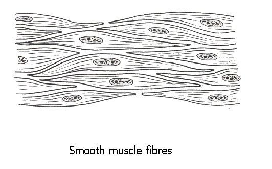
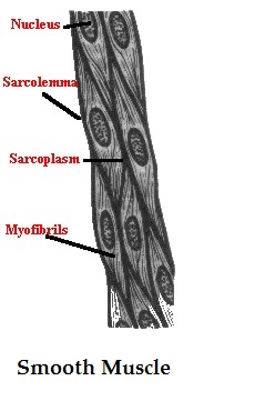
Smooth muscle tissue is made up of thin-elongated muscle cells, fibres.
These fibres are pointed at their ends
each has a single, large, oval nucleus.
Each cell is filled with a specialised cytoplasm, the sarcoplasm
Surrounded by a thin cell membrane, the sarcolemma.
Each cell has many myofibrils which lie parallel to one another in the direction of the long axis of the cell.
They are not arranged in a definite striped (striated) pattern, as in skeletal muscles
Hence the name smooth muscle .
Smooth muscle fibres interlace to form sheets or layers of muscle tissue rather than bundles.
Involuntary tissue, i.e. it is not controlled by the brain.
Smooth muscle forms the muscle layers in the walls of hollow organs such as the digestive tract - the walls of the bladder, the uterus, various ducts of glands and the walls of blood vessels .
Functions
Smooth muscle controls slow, involuntary movements such as the contraction of the smooth muscle tissue in the walls of the stomach and intestines.
The muscle of the arteries contracts and relaxes to regulate the blood pressure and the flow of blood.
Skeletal Muscle Tissue
Most abundant tissue
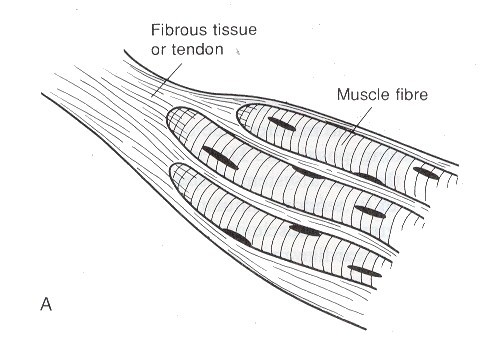
These muscles are attached to and bring about the movement of the various bones of the skeleton
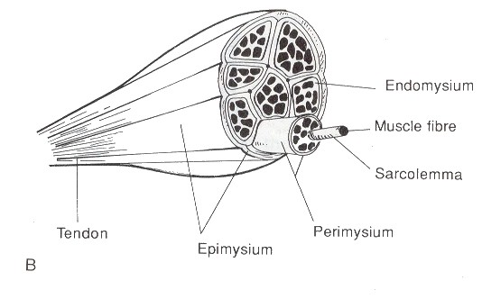
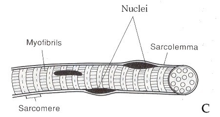
The whole muscle, such as the biceps, is enclosed in a sheath of connective tissue, the epimysium.
This sheath folds inwards into the substance of the muscle to surround a large number of smaller bundles, the fasciculi.
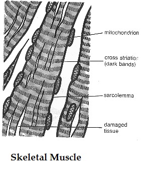
These fasciculi consist of still smaller bundles of elongated, cylindrical muscle cells, the fibres.
Each fibre is a syncytium, i.e. a cell that have many nuclei.
The nuclei are found at the periphery of the cell, just beneath the thin, elastic membrane (sarcolemma).
The sarcoplasm also has many alternating light and dark bands, giving the fibre a striped or striated appearance
Each muscle fibre is made up of many smaller units, the myofibrils.
Each myofibril consists of small protein filaments, known as actin and myosin filaments.
The myosin filaments are slightly thicker ( A-band).
The actin filaments are light bands (I-bands)
The actin filaments are attached to the Z-line.
This arrangement of actin and myosin filaments is known as a sarcomere.
During the contraction of skeletal muscle tissue, the actin filaments slide inwards between the myosin filaments.
Mitochondria provide the energy
This action causes a shortening of the sacromeres (Z-lines move closer together), which in turn causes the whole muscle fibre to contract.
This can bring about a shortening of the entire muscle
The contraction of skeletal muscle tissue is very quick and forceful.
Functions
Skeletal muscles function in pairs to bring about the co-ordinated movements of the limbs, trunk, jaws, eyeballs, etc.
Skeletal muscles are directly involved in the breathing process.
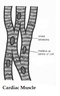
Cardiac Muscle Tissue
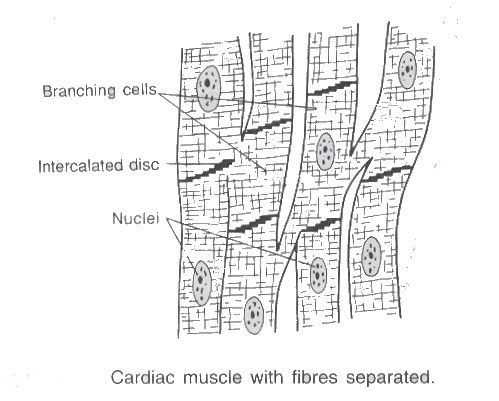
Found only in the walls of the heart.
Have cross-striations and contain numerous nuclei. The striations are not so obvious,
Involuntary.
The sarcolemma is thinner and not clearly discernible,
There is only one nucleus present in the centre of each cardiac fibre
Adjacent fibres branch and are linked to each other by so-called muscle bridges.
The spaces between different fibres are filled with areolar connective tissue which contains blood capillaries.
Functions
Cardiac muscle tissue plays the most important role in the contraction of the atria and ventricles of the heart.
It causes the rhythmical beating of the heart, circulating the blood and its contents throughout the body as a consequence.